


Arthropathy refers to a pathological process characterized by malnutrition and degeneration of articular cartilage. Usually, the problem is not limited to cartilage-the pathology later spreads to the bone tissue located under the cartilage (subchondral). Therefore, joint disease is also called osteoarthritis. Since all of these diseases eventually lead to changes in the structure of the joints, this process is called osteoarthritis deformans, and it can affect any joint. In clinical practice, arthropathy of the knee joint or knee joint disease will be noticed in most cases.
The nature of pathology
In terms of frequency and prevalence, knee arthropathy is second only to hip arthropathy (hip arthropathy). In order to find out the reasons for this situation, it is worth briefly introducing the characteristics of the knee joint anatomy and the functions it performs. This is one of the largest joints, which involves 3 bones-femur, tibia and patella. Therefore, it is a complex joint formed by 2 joints-the patellofemoral joint and the patellofemoral joint.
The articular surfaces of all 3 bones are covered with cartilage, which helps the movement of the joints and protects the subchondral bone tissue from mechanical wear. In addition to the articular cartilage itself, the knee joint also has a paired cartilage structure of meniscus, which can enhance the consistency of the articular surface (anatomical correspondence). Articular cartilage does not have its own blood vessels. Its nutrition is carried out by diffusion from the joint (synovial fluid). Like a sponge, cartilage shrinks under mechanical stress during exercise to carry heavy objects. At this time, waste is released from the cartilage tissue into the surrounding synovial fluid. On the contrary, at the moment of relaxation and rest, synovial fluid and its nutrients will penetrate into the cartilage of the knee joint.
Due to many reasons, the nutrition of the knee joint cartilage is disturbed, which leads to knee joint disease. At the same time, there was a lack of nutrients in cartilage tissue at first, such as chondroitin sulfate, glucosamine, calcium and other trace elements. Water loss. This is a process of malnutrition, followed by degradation-the thinning of articular cartilage. In turn, these negative processes can lead to structural and movement disorders of the knee joint.
Knee joint disease is often mistaken for salt deposits. For example, some mineral salts, including table salt, are deposited in the joint cavity in the form of microcrystals, which can cause pain and movement disorders. This is not true. Obviously, salt deposition takes a completely different process. In response to the destruction of articular cartilage in the subchondral bone, marginal bone growth-osteophytes-is formed to stabilize the knee at least to some extent. However, in the future, osteophytes will only aggravate joint disease and cause further destruction of cartilage.
reason
The causes of knee joint arthropathy are diverse, which may be related to the pathology of the knee joint itself, or may be related to other diseases and metabolic disorders. In this regard, knee joint disease can be primary and secondary. The mechanism of primary joint disease is not fully understood. It is believed that in this case, the disease is caused by a variety of factors, including:
- At advanced age, degenerative changes not only occur in articular cartilage, but also occur in all organs and tissues;
- Being overweight will increase the mechanical stress of the joints;
- Insufficient physical activity, and vice versa, excessive physical activity;
- Some congenital anatomical disorders of the knee joint, in which the articular cartilage and subchondral bone are initially changed;
- General metabolic disorders lead to changes in the mineral composition of synovial fluid.
Secondary arthropathy of the knee joint is a complication of other diseases. In most cases, this type of disease is arthritis of various natures-gout, rheumatism, rheumatoid, septic, tuberculosis, etc. In these diseases, various pathological factors (infection, abnormal immune response, uric acid crystals) form synovial inflammation in the so-called form. Synovitis. Synovitis inevitably accompanies the deterioration of synovial fluid quality, which in turn leads to arthropathy.
Another common cause of joint disease is knee injuries. Post-traumatic knee arthropathy is the result of intra-articular fractures of the femur and tibia, hemorrhage (arthropathy), knee ligaments and meniscus injuries. Here, pathology is based on mechanical factors (injuries) and subsequent injuries (arthritis). In addition, osteoporosis is often accompanied by arthropathy. The lack of calcium in the bones will not only destroy the bones, but also the cartilage tissue.
symptom
The main symptoms of knee arthropathy:
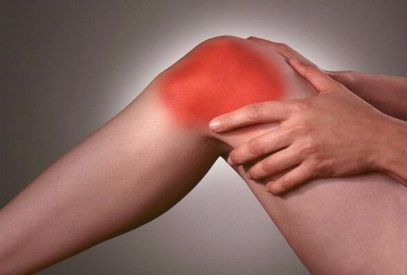
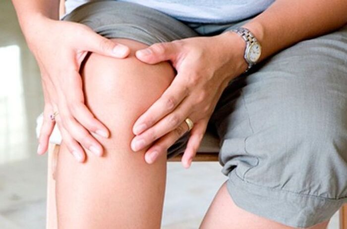
- pain;
- Impaired knee movement;
- Difficulty walking
- There is a crackling sound when moving;
- First-pathological tension, then-muscle atrophy of the lower limbs;
- Deformity of the knee joint.
At first, usually, the patellofemoral joint is affected, which accounts for most of the functional load. Generally speaking, knee pads with joint disease may be the most vulnerable. The dystrophic changes of arthropathy start from the cartilage of the knee. Clinically, this is manifested as swelling and pain when the bone is touched. As a result of changes in malnutrition, articular cartilage undergoes hardening changes-it loses its elasticity and is replaced by rough connective tissue.
Subsequently, the joint pockets and ligament devices undergo hardening changes. The configuration of the joints changes. Initially, it will swell and become inflamed due to the accompanying arthritis. Subsequently, with the progress of degeneration and hardening, the amount of synovial fluid decreases sharply, and the joint space becomes narrow, which inevitably leads to dyskinesia. At first, the gait is difficult and the muscles of the limbs are tense. Then rigidity develops-the knee does not move at all, and the result is atrophy of the thigh and calf muscles. All these changes are formed over a long period of time. In this regard, there are 3 degree arthropathy:
- 1 degree knee joint arthropathy. The pain is mainly concentrated in the kneecap area and the inner surface of the knee joint. Pain has an "onset" nature-they appear at the beginning of the exercise and then subside. In addition, pain occurs during strenuous exercise (long-distance walking, weight bearing), and it disappears after a rest. At this stage, there is no structural change in the joints.
- Second degree knee joint arthropathy. Pain occurs even when resting, and it lasts longer. The range of motion of the knee joint is limited (contracture). The patient limped and had to walk around with a stick. Formation of joint inflammation and malnutrition changes, externally manifested as an increase in knee joints due to edema.
- Third degree knee arthropathy. Severe knee pain that does not stop even if you rest for a long time. Severe and irreversible disorders of the joint structure, leading to joint stiffness and loss of athletic ability. The changes in the shape of the entire lower limbs are manifested as valgus or varus (O-shaped or X-shaped) bending.
The diagnosis of knee joint disease is based on the above symptoms and complaints of the patient and X-ray data (narrowing of the joint space, osteophytes, osteoporosis, bone sclerosis). Knee joint disease is treated through a combination of drugs and physical procedures. For third-degree arthropathy, surgical intervention is required, during which various types of knee joint plastic surgery are performed.